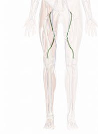
The Femoral Artery
Explore the anatomy, function, and role of the femoral artery with Innerbody's interactive 3D model.

The femoral artery is the main artery that provides oxygenated blood to the tissues of the leg. It passes through the deep tissues of the femoral (or thigh) region of the leg parallel to the femur.
The common femoral artery is the largest artery found in the femoral region of the body. It begins as a continuation of the external iliac artery at the inguinal ligament that serves as the dividing line between the pelvis and the leg. From the inguinal ligament, the femoral artery follows the medial side of the head and neck of the femur inferiorly and laterally before splitting into the deep femoral artery and the superficial femoral artery.
The superficial femoral artery flexes to follow the femur inferiorly and medially. At its distal end, it flexes again and descends posterior to the femur before forming the popliteal artery of the posterior knee and continuing on into the lower leg and foot. Several smaller arteries branch off from the superficial femoral artery to provide blood to the skin and superficial muscles of the thigh.
The deep femoral artery follows the same path as the superficial branch, but follows a deeper path through the tissues of the thigh, closer to the femur. It branches off into the lateral and medial circumflex arteries and the perforating arteries that wrap around the femur and deliver blood to the femur and deep muscles of the thigh. Unlike the superficial femoral artery, none of the branches of the deep femoral artery continue into the lower leg or foot.
Looking at the femoral artery in cross-section, it is made of several distinct tissue layers that help it to deliver blood to the tissues of the leg. The innermost layer, known as the endothelium or tunica intima, is made of thin, simple squamous epithelium that holds the blood inside the hollow lumen of the blood vessel and prevents platelets from sticking to the surface and forming blood clots. Surrounding the tunica intima is a thicker middle layer of connective tissues known as the tunica media. The tunica media contains many elastic and collagen fibers that give the femoral artery its strength and elasticity to withstand the force of blood pressure inside the vessel. Visceral muscle in the tunica media may contract or relax to help regulate the amount of blood flow. Finally, the tunica externa is the outermost layer of the femoral artery that contains many collagen fibers to reinforce the artery and anchor it to the surrounding tissues so that it remains stationary.
The femoral artery is classified as an elastic artery, meaning that it contains many elastic fibers that allow it to stretch in response to blood pressure. Every contraction of the heart causes a sudden increase in the blood pressure in the femoral artery, and the artery wall expands to accommodate the blood. This property allows the femoral artery to be used to detect a person's pulse through the skin.


