Pelvis

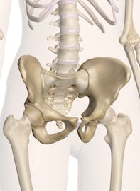
Located in the lower torso, the pelvis is a sturdy ring of bones that protects the delicate organs of the abdominopelvic cavity while anchoring the powerful muscles of the hip, thigh, and abdomen. Several bones unite to form the pelvis, including the sacrum, coccyx (tail bone), and the left and right coxal (hip) bones.
Throughout childhood, the pelvis is made of many smaller bones that eventually fuse during adulthood to form a more rigid pelvis. Each of the coxal bones begins as three separate bones: the ilium, ischium, and pubis. The ilium is the largest, widest, and most superior of the hip bones. When you place your hands on your hips, you can feel the curved ridge of the ilium known as the iliac crest. The narrow ischium is inferior to the ilium and is the bone, along with the coccyx, that you rest your body weight on while sitting. Anterior to the ischium is the pubis, the smallest of the hip bones. The ilium, ischium, and pubis meet in the center of the hip bone to form the deep, cup-like socket of the hip joint called the acetabulum.
The sacrum and coccyx also begin life as multiple bones before fusing. Five short, wide vertebrae fuse to form the wedge-shaped sacrum, while four tiny vertebrae fuse to form the coccyx.
The bones of the adult pelvis join together to form four joints: the left and right sacroiliac joints, the sacrococcygeal joint, and the pubic symphysis.
- The sacroiliac joints form between the sacrum and the left and right ilium to form a tight junction capable of supporting the body's weight and resisting the force of strong muscles. Although the sacroiliac joints are synovial joints, many strong ligaments and bony ridges on the sacrum and ilium interlock to prevent movement of the bones and strengthen the joint.
- The cartilaginous sacrococcygeal joint unites the sacrum with the tiny coccyx and allows some slight movement of the tail bone.
- On the anterior side of the pelvis the pubic symphysis unites the left and right pubic bones. The pubic symphysis is a slightly flexible band of fibrocartilage that allows the independent movement of the hip bones while walking. Women have a significantly wider and more flexible pubic symphysis compared to men, which allows the female pelvis to stretch during childbirth to accommodate the head of a fetus passing through the birth canal.
Many significant structural differences, or sexual dimorphisms, exist between the male and female pelvis. Most of these differences relate to the female reproductive system and the roles of pregnancy and childbirth in the female. The bones of the male pelvis are larger, thicker, and heavier than those of the female and there is very little hollow space within the ring of the pelvic bones. The female pelvis by comparison is significantly shorter and wider, which provides a greater hollow space within for the head of a fetus to pass through during childbirth. The sacrum and coccyx curve much more but are located more posteriorly in the female pelvis to increase the space of the pelvis. Even the shape and angle of the acetabulum and hip joint differ between males and females. The female acetabulum is rotated more anteriorly than in males, resulting in differences in walking gait and posture between men and women.


