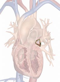
The Left Auricle
Explore Innerbody's 3D anatomical model of the left auricle, a vital component of pumping blood within the heart.

The left auricle, also known as the left atrial appendage (LAA), is a flap of heart wall on the anterior surface of the left atrium of the heart. Often confused with the left atrium, it is one of the most prominent structural features of the left atrium and plays an important role in the pumping of blood within the heart. The name auricle comes from the Latin word auricula, which means "ear" and refers to the floppy dog-ear shape of the auricle.
Anatomy
The left auricle is a thin pouch of the heart wall located on the anterior surface of the left atrium. Its walls are only about one sixteenth of an inch (1 mm) in thickness and less than 1 inch (2.5 cm) in length. The interior size of the left auricle changes considerably during each heartbeat, filling with blood and expanding before contracting to pump blood.
At its superior end, the left auricle merges with the walls of the left atrium. From this point, it descends inferiorly just anterior to the main chamber of the left atrium. At its inferior end, the left auricle has an irregular, scalloped shape with many tiny ridges. While the exterior of the auricle is quite smooth, its interior is filled with many tiny pockets, which allow it to hold a greater volume of blood when it expands.
Like all of the external parts of the heart, the walls of the auricle are made of three distinct layers of tissue.
- The outermost layer of the auricle is the epicardium, a layer of simple squamous epithelial tissue that gives the auricle a smooth, slick surface. Pericardial fluid is produced by the epicardium to lubricate its surface and prevent irritation during the movement of the heart.
- Deep to the epicardium is the myocardium layer that forms the bulk of the auricle's wall and contains cardiac muscle tissue. The myocardium produces the muscular contraction of the auricle, which allows it to pump blood.
- Deep to the myocardium is the endocardium, which forms the innermost layer of the heart. Endocardium is made of a specialized type of simple squamous epithelial tissue known as endothelium. Endothelium lines the inner surface of all blood vessels and the heart to keep blood cells within the circulatory system and to prevent platelets from sticking to the walls and forming blood clots.
Physiology
During the contraction cycle of the heart, oxygenated blood from the lungs passes through the pulmonary veins and enters the heart at the left atrium. The left atrium's job is to collect this blood and pump it to the left ventricle, which in turn pumps the blood to the body's tissues. While the left atrium has a large hollow space for this blood to collect, the left auricle is able to increase the collecting and pumping capacity of the left atrium without greatly increasing the size of the atrium. As blood fills the left auricle, it expands and holds the blood until the sinoatrial node of the heart sends a signal to the left atrium to begin contraction. At this point, cardiac muscle tissue in the myocardium contracts and squeezes blood out of the auricle and into the main cavity of the left atrium.
The left atrium continues to contract and pumps blood through the bicuspid valve to fill the left ventricle. While the left ventricle contracts, the bicuspid valve shuts to prevent regurgitation of blood. Meanwhile, new blood from the lungs fills the left atrium and left auricle again for the next round of pumping. The myocardium in the left atrium and left auricle relaxes, providing space for blood to fill their chambers again.


