The Anterior Longitudinal Ligament
Explore Innerbody's 3D anatomical model of the anterior longitudinal ligament, a vital component of the spinal column.

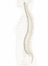
The anterior longitudinal ligament is a vital structural component of the spinal column. It connects all vertebrae and their intervertebral discs on the anterior side of the spine. Sudden movements in the neck can strain or tear the anterior longitudinal ligament, leading to the condition commonly known as whiplash.
Anatomy
The anterior longitudinal ligament is a long ligament found in the anterior of the spinal column. It arises from the inferior end of the anterior atlantoaxial ligament on the anterior surface of the vertebral body of the axis, or the C2 vertebra. From its origin, it descends along the spinal column, connecting the vertebral bodies of all the vertebrae from the axis to the sacrum and all of the intervertebral disks.
Like all ligaments, the anterior longitudinal ligament is made of dense regular connective tissue. The collagen fibers that make up this tissue are arranged in parallel bundles running vertically along the spinal column. In the anterior longitudinal ligament, the collagen fibers are divided into several distinct layers. The deep fibers are the shortest and closest to the bones, connecting one vertebra to its neighboring intervertebral discs and vertebrae. A middle band of intermediate fibers runs over the deep fibers and connects two to three vertebrae together. The superficial layer of fibers contains the longest fibers, which connect up to five vertebrae.
Physiology
The spinal column is a vital component of the skeletal system, which provides a strong yet flexible structure to the neck and torso. Many muscles and ligaments surround the bones of the spinal column to help support this vital structure. The anterior longitudinal ligament holds all of the bones of the spinal column and their intervertebral discs in alignment on the anterior side of the spinal column. It is slightly loose in its connection to the vertebrae, allowing a small range of movements between the bones in all directions.
Despite its strength, sudden, strong movements of the spine can damage the anterior longitudinal ligament, resulting in pain and stiffness in the neck. The most common occurrence of this movement is cervical acceleration/deceleration (CAD) syndrome, also known as whiplash. During whiplash, a sudden change in speed displaces the head and neck relative to the trunk in such a violent manner that the anterior longitudinal ligament is partially torn (sprained) or completely torn. Many activities may result in whiplash, but sports-related collisions and automobile accidents are the most common.

Copyright © Innerbody Research 1997 - 2025. All Rights Reserved.
