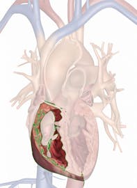
Right Ventricle

The right ventricle is second largest chamber of the heart, smaller than only the left ventricle. Like the left ventricle, it is a hollow, muscular chamber on the inferior end of the heart. The right ventricle performs the vital role in pulmonary circulation of pumping deoxygenated blood to the lungs where gas exchange occurs.
Anatomy
The right ventricle is located on the right inferior end of the heart. It extends superiorly from the apex of the heart to the right atrium. Its left side meets the left ventricle at the interventricular septum. Compared to the other chambers of the heart, the right ventricle is much larger and more muscular than the left and right atria, but is considerably smaller and less muscular than the left ventricle.
Two valves control the flow of blood through the right ventricle. The tricuspid valve connects the right atrium to the right ventricle and controls the flow of blood into the right ventricle. The pulmonary valve connects the right ventricle to the pulmonary trunk and controls the flow of blood exiting the right ventricle. Many thin chordae tendineae from the tricuspid valve extend into the right ventricle and connect to the papillary muscles in the walls of the right ventricle.
Histology
The visceral layer of the pericardium, also known as the epicardium, covers the walls of the right ventricle externally. This layer of simple squamous epithelium protects and underlying tissue and gives the right ventricle a smooth outer surface. Deep to the epicardium is the myocardium layer that makes up the bulk of the heart wall and contains the cardiac muscle tissue responsible for the pumping action of the heart. The innermost layer of the heart wall is the endocardium, a layer of simple squamous epithelial tissue that lines the heart. The endocardium prevents blood cells from sticking to the walls of the right ventricle and forming blood clots.
Physiology
Blood enters the right ventricle from the right atrium during ventricular diastole (relaxation) and atrial systole (contraction). Once the ventricle fills with blood, Purkinje fibers stimulate the cardiac muscle cells in the myocardium to contract, pushing the blood out of the right ventricle. Pressure from within the right ventricle closes the cusps of the tricuspid valve and prevents regurgitation of blood back into the right atrium. The right ventricle pumps blood through the open pulmonary valve into the pulmonary trunk, where it will continue on to the lungs before returning to the left side of the heart. At the end of ventricular systole, blood pressure in the pulmonary trunk closes the cusps of the pulmonary valve to prevent the regurgitation of blood back into the right ventricle. The tricuspid valve simultaneously reopens to permit the next batch of blood to be pumped into the right ventricle.


